Ankle Cartilage Injury
What is an Ankle Cartilage Injury?
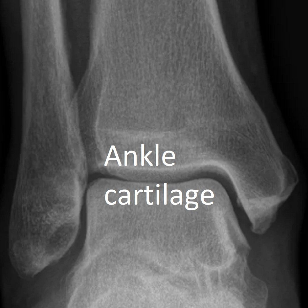
The ankle joint surfaces are covered by a smooth white layer of cartilage. This thin but very resilient tissue helps cushion the ankle and allows it to move smoothly. It is the "gap" seen between bone on X-rays.
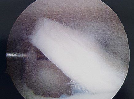
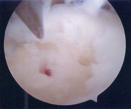
See X-ray and arthroscopic images below.
There are no nerves to detect pain in cartilage but the underlying bone and adjacent tissues have many. Cartilage has a poor blood supply and doesn't heal well.
Ankle cartilage injuries are also known as osteo-chondral injuries
because they often affect the underlying bone.
Ankle cartilage injuries are a common reason for ongoing ankle pain after a significant ankle sprain.
How do Ankle Cartilage Injuries Occur?
When the ankle joint is injured (sprained), the cartilage can be damaged in two ways:
- direct injury (common)
- injury to the underlying bone's blood supply (uncommon).
Symptoms of Ankle Cartilage Injury
The symptoms of ankle cartilage injury include:
- Pain and swelling that persists more than two months after an ankle sprain. Pain often feels "deep in the ankle"
- Clicking, jamming or locking of the ankle.
How is Ankle Cartilage Injury Diagnosed?
The diagnosis of ankle cartilage injury involves:
- taking a complete history
- performing a physical examination
- investigations.
These may include:
- weight-bearing X-rays of the ankle
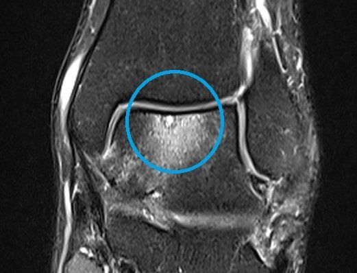
- CT and MRI (see image) of the ankle
- ankle arthroscopy.
How is Ankle Cartilage Injury Treated?
Non-surgical treatment
Unless a cartilage fragment has become loose in the ankle joint, non-surgical treatment is reasonable. Some can settle with time and protection.
Reducing impact activities is important. This means bike, gym and pool-based exercises rather than running. Shoes need to be supportive with heel cushioning.
Surgery for ankle cartilage injury
Surgery is indicated when a loose fragment is moving around the ankle joint or non-surgical treatment is unable to relieve ankle pain and swelling.
Surgery involves ankle arthroscopy and cartilage treatment. Depending on the size and depth of cartilage damage, options include:
- cartilage debridement and micro-fracture (+/- application of hydrogel bioscaffold) to stimulate healing (see images below)
- screw fixation of large osteo-chondral fragments (uncommon)
- transfer of cartilage from the knee to the ankle (OATS-mosaicplasty, uncommon)
- bone-grafting (uncommon)
- open surgery to access the inside of the ankle (medial malleolar osteotomy, uncommon).
If there is associated ankle instability, this is usually treated at the same time.
In most cases, recovery involves rest, gentle movements and crutches for two weeks, then two months of reduced impact activities. Final recovery is after six months.
More than 80% of people are helped by this surgery but recurrence is possible.
Please see the TREATMENTS menu for more information on Ankle Arthroscopy.


